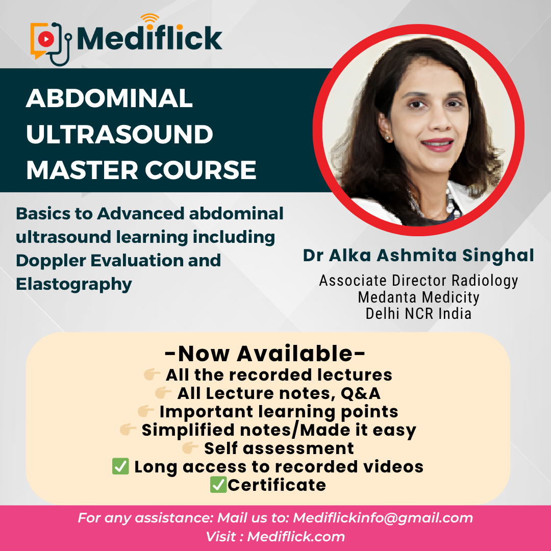
'); Abdominal Ultrasound Master Course
Contact us
Abdominal Ultrasound Master Course cover

Abdominal Ultrasound Master Course

Instructor: Dr. Alka Ashmita Singhal

Language: English

Valid Till: 2025-11-30

$281.09 37% OFF

$175.68 including 18% GST


ABDOMINAL ULTRASOUND MASTER COURSE

(Skill Perfection Masterclass series in Abdominal ultrasound including elastography and Doppler )


12 Modules with in-depth learning


Modules twice a month on Wednesdays

7-9 pm IST


Course Start Date: 18th December 2024, Wednesday


Notes with Q and A


Self-Assessment


Pre-session suggested reading


Faculty:

Dr Alka Singhal

Delhi NCR India


Introduction to course

This comprehensive Abdominal ultrasound master course is your one stop solution to abdominal ultrasound learning, starting from basics knobology, artefacts, transducer selection, presets, ultrasound technique and image optimisation, basics of Doppler principles, Practical skills, implementation and interpretation. The course includes understanding of Advanced modalities of elastography and CEUS. It covers all the essential abdominal organs in great detail. Dedicated modules are assigned for liver chronic liver disease, portal hypertension, cirrhosis, elastography, doppler, hepatic infections and benign and malignant tumours, and post-transplant liver evaluation. Common and uncommon pathologies of gall bladder and hepatobiliary tract, spleen, pancreas are included in dedicated modules. A special module covers the ultrasound evaluation of the gastrointestinal tract, acute appendicitis, intussesption and other common ultrasound pathologies in paediatric and adult age group. The final modules cover the genitourinary tract , with a dedicated module for kidneys and urinary tract, followed by gynaecology modules covering the uterine ovarian and adnexal pathologies.

Come one ! Come All ! Come with all your inquisitiveness to learn, and enhance your skills, and with all your questions. Interactive QA session is available at the end of each module.


Tentative Scientific Schedule

Date and Time

ABDOMINAL ULTRASOUND MASTER COURSE


Module -1
18th December 2024 (7-9 pm)
Wednesday

Basics of Ultrasound, Colour Doppler and Elastography

Module – 2
8th January 2025 

(7-9 pm)
Wednesday

Ultrasound of Liver – Normal, Congenital and infective diseases.


Module -3
29th January 2025 (7-9 pm)
Wednesday

Ultrasound of Liver – Chronic liver diseases and Colour Doppler

Module – 4
12th February 2025  (7-9 pm)
Wednesday

Ultrasound of Liver – Neoplasm and liver transplant

Module – 5

26th February 2025
(7-9 pm)
Wednesday

Spleen, Biliary tract, gall bladder and pancreas

Module – 6

12th March 2025
(7-9 pm)
Wednesday

Gastrointestinal tract

Module – 7

26th March 2025
(7-9 pm)
Wednesday

Kidney and Urinary Tract: Part- 1

Module – 8

9th April 2025
(7-9 pm)
Wednesday

Kidney and Urinary Tract: Part -2

Module – 9 
23rd April 2025

(7-9 pm)
Wednesday

Uterus

Module -10 
7th May 2025 

(7-9 pm)
Wednesday

Ovaries and fallopian tubes

Module -11
21st May 2025

(7-9 pm)
Wednesday

Anterior Abdominal Wall Ultrasound Evaluation: and Anterior Abdominal Wall Hernias

Module -12
4th June 2025

(7-9 pm)
Wednesday

Ultrasound Scrotum


Frequently asked question

1. How to Join/ How to access recording of lectures

Ans: After successful purchase, this course will be added to your courses.

You can access Live session/ recording in the following ways:

  • From Web browser -
    • After successful login, go to the “My courses” Section (just left to the login in the right upper corner) and click the course and watch from there.
  • Android / iOS App
    • Download the Android / iOS app and after a successful login go to “Library” in the lower bar and click the course and watch from there.

For other devices, you can access courses through the web browser of your device.

Kindly note: Join the Whatsapp group of live course/conference after the registration for better communication and to remain updated.

We also give some special discount for participant of the concern course in future courses, that too updated in whatsapp group and in Email


2. Will the course link be sent to us on E-mail or whatsapp?

Ans:  No direct link will be sent to your E-mail or Whatsapp.

Though we will send you reminder Email and Whatsapp message to join live session.
To get E- mail reminder mark Mediflickinfo@gmail as non-spam, otherwise Email reminder may go to spam folder and you may not be aware of that.


3. I forgot the password to log in on Mediflick.com, what to do?
Ans: Just reset your password. You will get password reset mail. In case you don’t find password reset mail in your inbox, check in spam folder


4. I am unable to log in. I get this message stating I can access only from 10 devices, what to do?

 Ans:  Log in on every browser or app is considered one device so try to log out from other browser. If issue still persist then kindly E-mail to us at Mediflickinfo@gmail, we will manually reset no. of devices in 1-2 working days usually.


5. When I will get my certificate of completion of the course/ conference?

 Ans:  You can manually download your certificate after the completion of the course

For conference, We manually Email certificate after few days of conference


6. When I will get recording of the live course if available?

 Ans:  Usually it takes 24-48 hours to access recording after the live course. But in case of any technical issue it may take some longer time. Duration of access to recording is counted after it’s available for participants.


7. I could not complete my course due to some reason, is it possible to get extended access of the recording?
 Ans:  It’s not possible to extend the recording after it ends. You should purchase longer duration access option in the course if available or you may have to repurchase the course.

In case of any further question or if you feel any issue kindly write to us and also send us screenshot or video of the issue on Mediflickinfo@gmail.com 

Keep Learning
Mediflick.com


Details of the Modules

Module 1 - Basics of Ultrasound, Colour Doppler and Elastography

graphy

  • Ultrasound Physics, knobology, artefacts, transducer selection, presets, ultrasound technique and image optimisation.  
  • Basics of Doppler principles, Practical skills, implementation and interpretation.  
  • Understanding of Advanced modalities of elastography and CEUS (Contrast enhanced ultrasound). Bioeffects of ultrasound Imaging.  


Module 2 – Ultrasound of Liver – Normal, Congenital and infective diseases.

  • Liver: Normal ultrasound appearances; size and echotexture assessment. Segmental Anatomy and identification on ultrasound.   
  • Congenital abnormalities : Hepatic cysts, peribiliary cysts, polycystic hepatic disease, biliary hamartoma.  
  • Infectious diseases: Viral Hepatitis, Hepatic Abscess- Pyogenic and fungal.  Parasitic diseases: Amoebic liver Abscesses and hydatid disease

Module 3 - Ultrasound of Liver – Chronic liver diseases and Colour Doppler

  • Fattly Liver , NAFLD (Non alcoholic fatty liver disease), and Role of Elastography and Ultrasound Fat Quantification  
  • Hepatic cirrhosis and Portal hypertension  
  • Doppler evaluation in chronic liver disease, TIPS Shunt evaluation , PVT, Budd Chiari Syndrome  


Module 4 - Ultrasound of Liver – Neoplasm and liver transplant

  • Liver : Hepatic Masses: Benign and Malignant ;Detection and characterisation of focal hepatic lesions.
  • Benign hepatic neoplasms : Hemangioma (Typical and atypical, appearances, small and large lesions), FNH (Focal Nodular hyperplasia) Hepatic Adenoma, Hepatic Lipomas and angiomyolipomas  
  • HCC (Hepatocellular carcinomas) and metastatic liver disease.
  • Hepatic Trauma. Role of CEUS (Contrast enhanced ultrasound)  
  • Post liver transplant Doppler and Intraoperative hepatic ultrasound  


Module 5 – Spleen, Biliary tract, gall bladder and pancreas

  • Spleen : Sonoanatomy, situs inversus and identification of spleen, size and echotexture assessment.
  • Accessory spleen and splenenculus. Splenic cysts: simple and hydatid cysts. Splenic nodular lesions and tuberculosis of spleen, Gamma Gandy Bodies. Splenic microabscesses and large abscesses. Focal solid lesions: Hodgkins disease and Non Hodgkins lymphoma. Splenic Metastasis. Splenic Trauma  
  • The biliary tract: Normal anatomy: Choledochal cysts, carolis disease, evaluation of cholestatic jaundice. Choledocholithiasis and scanning tips for better demonstration and to avoid pitfalls. IHBRD (Intrahepatic biliary tract dilatation), Pneumobilia, Cholangitis, Cholangiocarcinoma  
  • Gall Bladder : Normal size range and wall assessment. Gall Bladder wall edema. Cholelithiasis (types and sizes) Biliary Sludge, Cholecystistis- Acute and Chronic Cholecystitis. Gall bladder perforation. Porcelain Gall bladder. Poypoidal masses of Gall Bladder: Cholesterol polyps, GB adenomas and malignancies.  

Pancreas : Sonoanatomy and technique of ultrasound evaluation. Normal parenchymal appearances and size. Assessment of pancreatic duct. Acute Pancreatis and evaluation of complications. Chronic Pancreatitis. The diagnostic spectrum. Pancreatic SOL -cystic and solid and pancreatic masses, Pancreatic Carcinoma. GEP-NET’s (Gastroenteropancreatic neuroendocrine tumors). Role of CEUS


Module 6 - Gastrointestinal tract

  • Role of ultrasound imaging in the evaluation of gastrointestinal tract: Anatomy and sonographic technique, Graded compression sonography: The Gut signature, Doppler evaluation of gut wall, CEUS and elastography of bowel.   
  • Acute Appendicitis: perforation of appendix, periappendiceal abscess, appendicolith, Mucocele of appendix   
  • Acute Diverticulitis and Acute typhlitis. Acute omental infarction. Inflammatory bowel disease: Crohn disease.  

Intussessption and hypertrophic pyloric stenosis. Hyperperistalsis and hypoperistalsis, Paralytic ileus. GIT Neoplasms and Lymphoma


Module 7 - Kidney and Urinary Tract: Part- 1

  • Kidney and Urinary Tract: Normal anatomy and sizes, Echotexture assessment, Corticomedullary differentiation.
  • Congenital anomalies: Ectopia, crossed renal ectopia, horseshoe kidney, Duplex collecting system and ureterocele.
  • Calcification : Renal calculi, ureteral calculi, Bladdder calculi. Nephrocalcinosis, Medullary nephrocalcinosis.
  • Hydronephrosis: PUJ Obs, congenital megacalyces, congenital megaureter and more..  
  • Renal cortical cysts and medullary cysts: Medullary sponge kidney, Polycystic kidney disease, Medical Renal Disease, AKI(Acute kidney injury), CKD (Chronic kidney disease )
  • Pyelonephritis, Pyonephrosis, Renal Papillary necrosis, Renal and Perinephric abscess, cystitis, pseudopolyp appearance in urinary bladder 


Module 8 - Kidney and Urinary Tract: Part -2

  • Genitourinary tumors: Angiomyolipoma, Renal cell carcinoma and other tumors, Transitional cell carcinoma, Lymphoma and metastasis. Adrenal lesions. Renal trauma.  
  • Urinary bladder diverticula and masses, Prostate, TRUS, Elastography. Special considerations for paediatric urinary tract  
  • Renal Arterial Doppler and evaluation of  Post Renal Transplant recipient.  


Module 9 – Uterus

  • TA and TVS (TVS is needed for optimal assessment), Sono HSG Scanning Techniques and normal uterus appearances at various ages. Congenital anomalies, Role of 3D  
  • Uterine enlargement:  fibroids and adenomyosis, leiomyosarcomas, ACUM, Intrauterine devices assessment.  
  • Normal endometrium appearances and endometrium pathologies. RPOC and AVM. Pelvic congestion syndrome.
  • Evaluation of uterine cervix.  


Module 10 - Adnexa : Ovaries and fallopian tubes

  • Techniques of assessment and normal ultrasound appearances at various age and with the phases of menstrual cycle. Follicular monitoring and assessment. Functional ovarian cysts, PCOD  
  • Benign ovarian lesions: Ovarian Endometriotic cysts, Ovarian Dermoid. Ovarian Torsion  
  • Ovarian Neoplasms: IOTA and ORADS. Metastatic tumors.
  • Fallopian tube abnormalities: Pelvic inflammatory disease. Tubal Ectopic gestation   


Module 11 - Anterior Abdominal Wall Ultrasound Evaluation: and Anterior Abdominal Wall Hernias

  • Diverification of Rectii , Inguinal canal sono anatomy and Inguinal Hernias. Indirect Inguinal Hernias,Direct Inguinal Hernias, Femoral Hernias, Spigelian Hernias, Sports Hernias
  • Complications of hernia. Evaluation in post surgical patient: Linea Alba Hernias, Umbilical Hernias, Paraumbilical or Periumbilical Hernias, Incisional Hernias, Multiple Hernias, Recurrent Groin Hernias, Hernia mimics
  • Lumps  and Bumps: Miscellaneous lumps, lipoma, scar endometriosis  


Module 12 : Ultrasound Scrotum

  • Sonographic technique, Normal anatomy
  • Benign Intratesticular Lesions, Cysts,  Tubular Ectasia of Rete Testis, Cystic Dysplasia, Epidermoid Cysts, Abscess, Torsion, Segmental Infarction, Calcifications  
  • Extratesticular pathologic Lesions: Tunica Vaginalis Hydrocele, Hematocele, and Pyocele, Paratesticular Masses, Varicocele
  • Epididymal Lesions, Acute and Chronic Epididymitis, Sarcoidosis
  • Fournier Gangrene, Trauma,   Cryptorchidism  
  • Malignant Tumors : Germ Cell Tumors, Non–Germ Cell Tumors, Testicular Metastases
  • Lymphoma and Leukemia, Extramedullary Myeloma  


Reviews
Other Courses
Launch your GraphyLaunch your Graphy
100K+ creators trust Graphy to teach online
𝕏
MEDIFLICK.COM. All Rights Reserved 2024 Privacy policy Terms of use Contact us Refund policy