There are no items in your cart
Add More
Add More
| Item Details | Price | ||
|---|---|---|---|

.png)
Instructor: Multiple Faculties
Language: English
Validity Period: 365 days
10% DISCOUNT – On Limited Seat
Starting On 11 October 2026
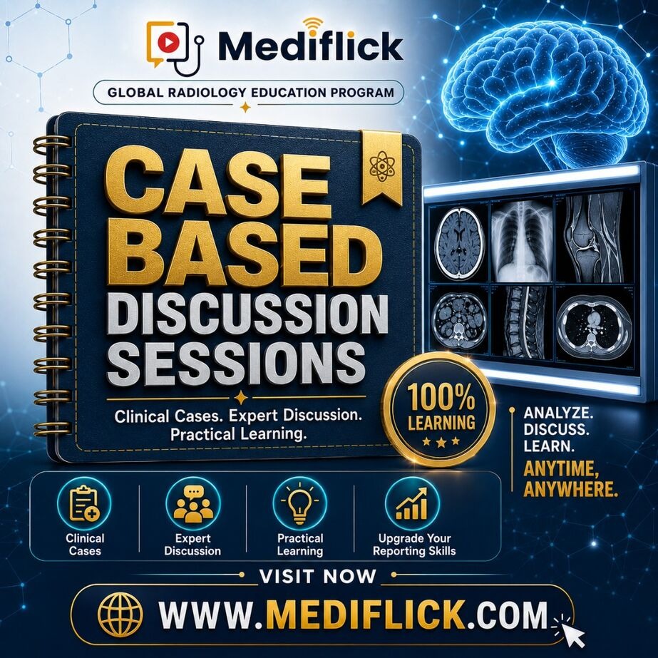
What you will get (Global Unique features) – 💪🏼Who else provide all this??????
⏱️ Duration: 1 Hour per Session
🎯 Total Academic Teaching: 24 Hours
🩻 Format: Case-Based • Interactive
📚 Coverage: Major Radiology Systems & Modalities
CASE → OBSERVATION → ANALYSIS → DIFFERENTIAL → DIAGNOSIS → REPORT
Classic cases. Frequently asked examination spotters. High-yield teaching points.
What will you get -
✘ No travel
✘ No accommodation
✘ No leave required
You may know a topic very well theoretically.
But when an actual CT, MRI, X-ray or ultrasound case appears, many questions arise:
👉🏼 Where should I start?
👉🏼 Which finding is important?
👉🏼 Is this finding primary or incidental?
👉🏼 What differential diagnosis should I consider?
👉🏼 Which diagnosis fits best?
👉🏼 What should I mention in the Reports?
👉🏼 What important finding should I not miss?
Case-based learning helps bridge the gap between:
By discussing cases step by step, participants will learn how to:
✅ Approach an unknown case systematically
✅ Identify key imaging abnormalities
✅ Build appropriate differential diagnoses
✅ Narrow the differential logically
✅ Recognize important imaging patterns
✅ Interpret findings in clinical context
✅ Avoid common diagnostic mistakes
✅ Identify important positive and negative findings
✅ Structure the final diagnosis
✅ Improve radiology reporting
✅ Answer examination and viva questions confidently
The cases will be approached in a structured manner.
Before jumping to a diagnosis:
• Which modality is being shown?
• Which organ/system is involved?
• What is abnormal?
• What is the dominant imaging finding?
• Is the abnormality focal, diffuse, unilateral or bilateral?
The abnormality will then be characterized systematically.
Depending on the case:
• Location
• Size
• Shape
• Margin
• Density / signal intensity
• Enhancement pattern
• Calcification
• Diffusion restriction
• Fat
• Hemorrhage
• Necrosis
• Vascularity
• Relationship with surrounding structures
• Associated findings
The discussion will focus on:
Participants will learn how to organize differentials based on:
• Age
• Clinical presentation
• Location
• Imaging morphology
• Signal/density characteristics
• Enhancement
• Associated findings
The most important question:
The discussion will focus on:
👉🏼 Diagnostic clues
👉🏼 Discriminating features
👉🏼 Classic imaging signs
👉🏼 Supporting findings
👉🏼 Findings arguing against competing diagnoses
Some of the most useful learning happens when a diagnosis is initially missed.
Important traps will be discussed, including:
❌ Jumping to the diagnosis too early
❌ Missing a subtle finding
❌ Anchoring on the clinical history
❌ Overcalling normal variants
❌ Confusing similar-looking pathologies
❌ Ignoring negative findings
❌ Giving unnecessarily broad differentials
❌ Missing an important associated abnormality
Whenever relevant, cases will move beyond diagnosis into reporting.
You will learn:
Including:
• Important descriptive findings
• Relevant measurements
• Important negative findings
• Complications
• Staging-related information
• Management-relevant findings
• Appropriate impression
• When to suggest additional imaging
Important cases may be followed by questions such as:
What is your diagnosis?
What are your differential diagnoses?
Which feature favours your diagnosis?
What is the classical imaging sign?
What is the best modality?
What additional sequence would you perform?
What complications should you look for?
What is the next step in management?
How would you report this case?
The 24 sessions will cover case-based discussions across the major areas of radiology.
These are not conventional lectures where the diagnosis is already written on the first slide.
The participant will be encouraged to think through the case.
CASE → OBSERVATION → ANALYSIS → DIFFERENTIAL → DIAGNOSIS → REPORT
Many cases will include real-world decision-making questions:
👉🏼 Would you advise contrast?
👉🏼 Which MRI sequence would you add?
👉🏼 Is ultrasound sufficient?
👉🏼 Should CT or MRI be performed next?
👉🏼 Is biopsy needed?
👉🏼 Is follow-up appropriate?
👉🏼 Does the finding require urgent communication?
This helps convert imaging knowledge into practical radiology.
❌ Looking only at the obvious lesion
❌ Missing a second abnormality
❌ Forgetting to review previous imaging
❌ Overdiagnosing incidental findings
❌ Giving too many differentials
❌ Failing to recognize emergencies
❌ Missing staging information
❌ Writing an impression that does not answer the clinical question
Where appropriate, cases will include:
✅ What should be described
✅ What should be measured
✅ What terminology should be used
✅ What findings matter for the clinician
✅ What should be mentioned in the impression
✅ How to avoid vague reporting
✅ How to make the report clinically useful
The goal is to gradually change the way you look at a case.
Instead of:
Move towards:
That is the difference between memorising radiology and learning radiology.
This programme is especially useful for:
✅ First-year radiology residents
✅ Second-year radiology residents
✅ Final-year MD/DNB residents
✅ Residents preparing for university examinations
✅ Post-MD/DNB candidates preparing for competitive examinations like FRCR,EdiR etc.
✅ Young radiologists looking for systematic revision
✅ Anyone wanting to improve case interpretation and reporting
The programme aims to help you become better at:
✅ 24 focused case-based sessions
✅ 24 hours of interactive radiology learning
✅ System-wise comprehensive coverage
✅ X-ray, ultrasound, CT, MRI and other modalities
✅ Common cases + important uncommon cases
✅ Diagnostic approach to unknown cases
✅ Differential diagnosis discussion
✅ Imaging signs and patterns
✅ Common traps and mistakes
✅ Reporting tips and pearls
✅ Examination-oriented discussion
✅ Viva-style questioning
✅ Practical day-to-day radiology relevance
No. The primary focus is case-based discussion and active image interpretation rather than conventional topic lectures.
Yes. The 24 sessions are planned to cover the major systems encountered during radiology residency.
Yes. Beginners can learn a systematic approach to cases, while senior residents can use the same cases for advanced differential diagnosis and examination preparation.
Yes. Wherever relevant, important reporting points, impression formulation and management-relevant findings will be discussed.
The emphasis will be on a balanced mix of common diagnoses, must-know cases, important emergencies, classic cases, examination favourites and selected uncommon conditions.
Yes. Although the primary goal is improving practical radiology reasoning, the format naturally supports preparation for viva, long cases, short cases and spotter-based examinations.
Cases may include X-ray, ultrasound, Doppler, CT, MRI, mammography and other relevant imaging modalities, depending on the system.
UNDERSTAND HOW TO REACH IT.**
Because in an examination—and in real-life reporting—the case in front of you may not look exactly like the one in the textbook.
Visit registration page for detailed SCIENTIFIC PROGRAM
📱 Mediflick available on Android & iOS for recorded learning ✅
💓 Join Mediflick Daily Learning Group (send intro to get added):
For any assistance:
📞 Helpdesk: +91 88906 81987
📧 Email : Mediflickinfo@gmail.com
